The History of Bedside Ultrasound: From Submarines to Sub-Interns
July 30, 2014
By Michael Vogel
Among the myriad of modern diagnostic tools, few can claim the certainty, consistency, and intimacy of ultrasound. In contrast to other dominant types of medical imaging characterized by large, foreign machines and uncomfortable noise and positioning, this sound-based imaging technique is one of the least intimidating and widely-used exam method, applied in fields ranging from Pulmonology and Gastroenterology to Obstetrics and Gynecology. However, conventional ultrasound can trace its roots to a form of militarized SONAR developed heavily in the period between World War I and II. Prototypes of “Ultrasonic Metal Flaw Detectors”, developed in tandem by Soviet scientist Sergei Y Sokolov and American engineer Floyd A Firestone in the early 1930s were used as an alternative to conventional RADAR methods for detecting submerged vehicles. These tools used short, high-frequency ultrasonic pulses to measure imperfections in the cast metal bodies of submarines, and their application in therapeutic medicine was simultaneously explored by American physicians. During a period stretching from the mid-1930s to the late- 1940s, ultrasound technology was used to perform a primitive form of craniotomy, by using high-frequency pulses to destroy parts of the brain. Additionally, this homeopathic application of ultrasound attempted to treat in patients with Parkinsonism, as well as other illnesses that include rheumatism, carcinomatosis, eczema, urinary incontinence, and elephantiasis. In fact, ultrasound wasn’t considered a diagnostic method until October of 1949 with the publishing of the “June ’49 Report” by George Ludwig. Since then, ultrasound has matured into “medical sonography”, and has expanded to become the primary diagnostic method for identifying the size, location, and pathology of muscles, tendons, and most internal organs.

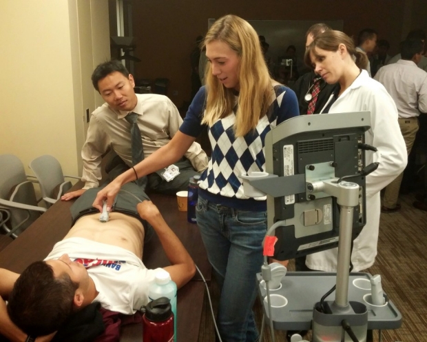
At Stanford Hospital, as well as many other prominent medical facilities worldwide, the use of bedside ultrasound is an essential tool in the repertoire of a hospitalist. Conventionally defined, Bedside Ultrasound refers to “a limited ultrasound examination performed at the bedside by the treating physician to answer specific clinical questions”. While bedside ultrasound originated in the quick decision-making of E.R. Physicians, it has now also found a home in internal medicine. Physicians use ultrasound to estimate central venous pressure through visualization of the inferior vena cava, estimate left ventricular function with limited echo, assess abnormalities in the lungs, evaluate the presence of ascites and pleural effusions, and to assist with procedures. As part of the physical exam, bedside ultrasound gives the opportunity to foster the physician-patient relationship. The Stanford Medicine 25 team providesinformative resources that explore the depth of ultrasound technique, from the proper placement of the transducer to motions and technique that maximize diagnostic efficiency while minimizing patient discomfort. Ultrasound technology, with its rich history and varied application remains today as one of the most widely-utilized assisted-examination techniques, and is a fantastic way to integrate imaging technology into a comprehensive and patient-forward physical exam.
P.S: Some facilities warm their ultrasound gel before applying it on a patient’s body. This action serves no diagnostic purpose and is purely for the patient’s comfort, a philosophy we could extend to our physical exams as well!
SOURCES
http://www.ob-ultrasound.net/history1.html
http://aquasonicultrasoundgel.com/blog/top-five-benefits-of-an-ultrasound-gel-warmer/
http://www.nlm.nih.gov/medlineplus/ency/article/003336.htm
http://www.slredultrasound.com/Filesandpictures/Guidelines2.pdf