Clinical Signs of Systemic Sclerosis
August 16, 2017
Christopher Barrett, MD
Case Presentation:
A 52 year old man presents to clinic with multiple alarming complaints. He is a Hispanic immigrant and has not received medical care in the United States. His primary complaints are dysphagia with frequent regurgitation, chronic melena, myalgias, pruritus, progressive fatigue and dyspnea on exertion, and painful swelling of his fingers with occasional discoloration in his fingertips.
1. What is his specific diagnosis?
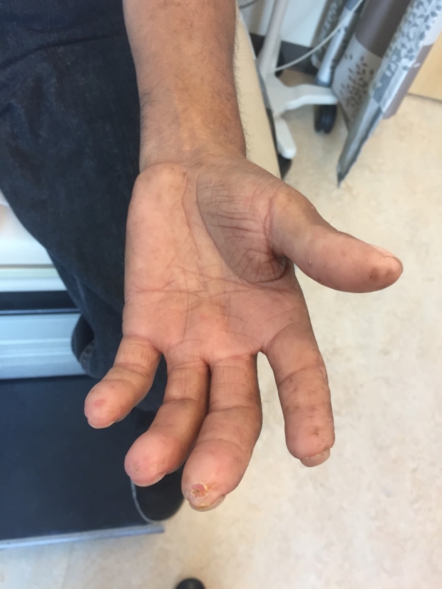
2. What are each of the hand findings depicted below?
.JPG)
Answer:
Examination of his hands reveals sclerodactyly, a thickening of the skin involving the full length of the digits. Skin thickening in this patient extends to the shoulders and chest. This is most consistent with a diagnosis of diffuse cutaneous systemic sclerosis, unlike limited cutaneous systemic sclerosis which normally only involves the fingers, toes, face, and distal extremities. This patient was diagnosed with diffuse cutaneous systemic sclerosis based on clinical findings and autoimmune serologies.
In the second panel, you can see that the patient has lost the digital pulp of his index finger and has evidence of digital pits on his fingertips. The third finger shows a loss of epithelialization consistent with a digital ulceration. All of these findings are caused by poor blood flow to the fingers. Though not specifically pictured here, patients frequently exhibit resorption of the distal phalanges, known as acro-osteolysis. This can look like bulbous enlargement of the digits due to clubbing, but is actually a distinct clinical entity with a different pathophysiology.
What is systemic sclerosis?
Scleroderma is an umbrella term for a group of autoimmune conditions which all demonstrate the common clinical finding of sclerotic (thickened) skin. Scleroderma may be localized, involving only the skin, or may be part of a more generalized condition known as systemic sclerosis. Patients with systemic sclerosis exhibit sclerotic changes of the skin, but also have evidence of damage to visceral organs as a result of their autoimmune disease. Systemic sclerosis can be further subdivided into limited cutaneous (lcSSc) and diffuse cutaneous systemic sclerosis (dcSSc). Both limited and diffuse cutaneous systemic sclerosis patients have systemic disease characterized by Raynaud’s phenomenon, the presence of autoantibodies, and internal organ involvement. Patients with limited cutaneous SSc usually have sclerosis only of the distal acral extremities and/or the face. They often present with a constellation of symptoms known as CREST syndrome (Calcinosis, Raynaud’s, Esophageal Dysmotility, Sclerodactyly, and Telangiectasias). Patients with diffuse cutaneous SSc present with axial as well as acral sclerosis. They may have all of the above CREST manifestations as well, but are also at high risk of developing significant pulmonary, renal, cardiac, musculoskeletal, and gastrointestinal manifestations of this disease.
What other exam findings can be seen in systemic sclerosis?
In some patients, cutaneous calcium deposits (calcinosis cutis) can be found on the hands and other parts of the acral appendages (see image below for example). This patient was also noted to have areas of skin hyper and hypopigmentation on his chest known as a salt & pepper rash. He has prominent skin thickening on his face, in particular over his eyebrows and around his lips, and he has telangiectasias scattered throughout his axial skeleton. Fine velcro crackles can be heard on examination of his lungs. He has diffuse muscle atrophy, bilateral knee effusions, and conjunctival pallor. Capillaroscopy was not performed in this clinic to evaluate for nail capillary loop dilation.
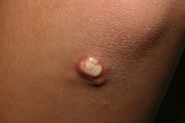
Calcinosis cutis - Calcium deposition in skin seen in systemic sclerosis.
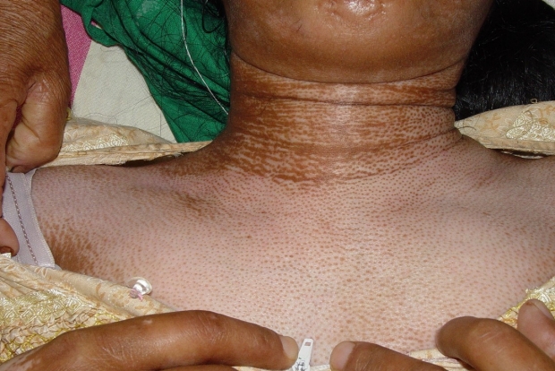
Salt & Pepper Skin - Hyper and hypopigmentattion seen in systemic sclerosis.
Courtesy of Vandana Mehta Rai MD, and C Balachandran MD via Derma tology Online Journal
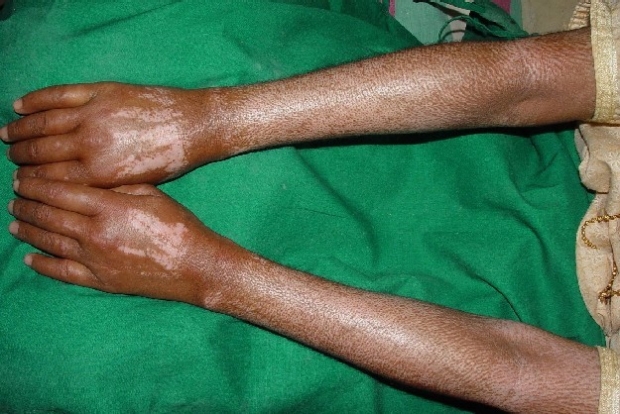
Salt & Pepper Rash of Arms - Hyper and hypopigmentattion seen in systemic sclerosis.
Courtesy of Vandana Mehta Rai MD, and C Balachandran MD via Derma tology Online Journal
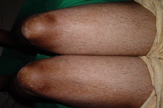
Salt & Pepper Rash of Thighs - Hyper and hypopigmentattion seen in systemic sclerosis.
Courtesy of Vandana Mehta Rai MD, and C Balachandran MD via Derma tology Online Journal
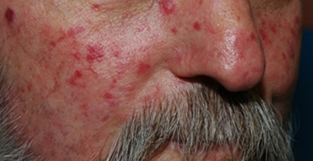
Matted telangiectasia common in scleroderma. Image courtesy of Dr. Lorinda Chung.
What is the typical workup for systemic sclerosis?
Some areas to focus on include the GI tract (esophageal dysmotility, gastric antral vascular ectasia, small bowel bacterial overgrowth), lungs (interstitial lung disease, pulmonary hypertension), joints, muscles, and routine labs (e.g. anemia). Patients with early diffuse disease are also at risk for scleroderma renal crisis. Further workup in the case of systemic sclerosis can often be guided by symptoms to evaluate for specific organ involvement.
This patient’s initial laboratory workup revealed strong positivity of serum auto-antibodies (ANA, Anti-Scl70), profoundly elevated CK and aldolase levels, and severe iron deficiency anemia. He had pulmonary function testing and high-resolution CT of his thorax to evaluate for pulmonary pathology due to his complaints of dyspnea and his abnormal respiratory exam. PFT’s showed a severe restrictive pattern, and his CT revealed interstitial fibrosis with prominent ground glass opacities in the lung bases. Echocardiogram did not reveal any evidence of pulmonary hypertension or cor pulmonale. His melena, fatigue, and iron deficiency anemia prompted evaluation for gastrointestinal bleed. Esophagogastroduodenoscopy (EGD) demonstrated reflux esophagitis, esophageal stricture, and vascular ectasias in the antrum of the stomach (GAVE).
What happened to our patient?
After diagnosis, this patient was referred to rheumatology subspecialty clinic where he could be evaluated and treated for diffuse cutaneous systemic sclerosis. He was started on immunosuppression with a TNF inhibitor, and will be closely monitored for progression of interstitial lung disease. He continues to take amlodipine for Raynaud’s, and hydroxyzine for symptomatic relief of his generalized pruritus. He underwent laser ablation of his gastral ectasias and was started on iron supplementation for anemia due to his chronic gastrointestinal bleeding.
Acknowledgement
Thank you to Dr. Lorinda Chung, Associate Professor of Medicine and Stanford Rheumatologist, for input on this this case.