Approach to the Low Back Exam
Low back pain is one of the most common complaints and most commonly caused by musculoskeletal issues. However it is important to know the exam so that you can confirm less worrisome musculoskeletal issues and look for more serious causes.
Introduction
Low Back Exam
Back pain is one of the most common complaints in the outpatient setting. While most causes are related to either the bone contacting the nerves as they exit the verbral canal or strain of the lumber muscles, it's important to be able to confirm this cause with the exam and know when more serious causes such as malignancy, infection (e.g. lumbar osteomyelitis) and inflammatory arthritis, to name a few.
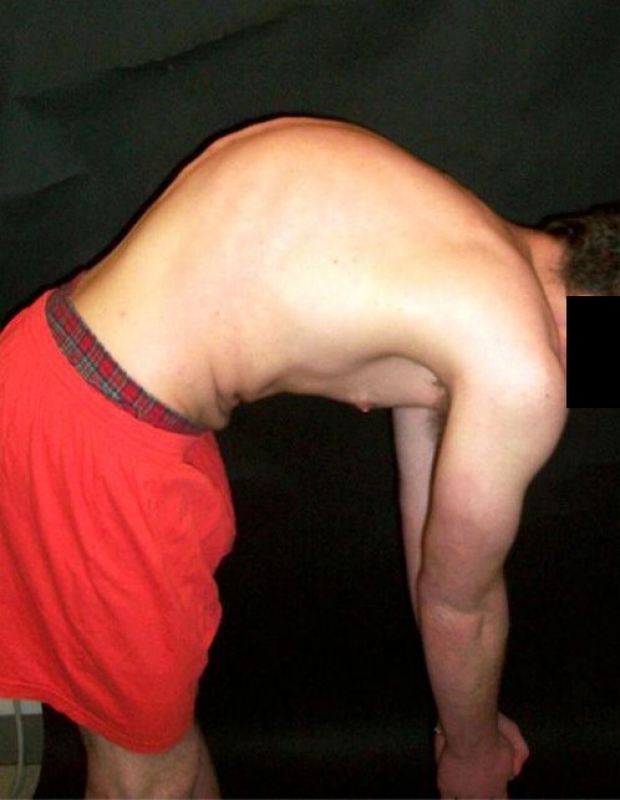
Inspection
Low Back Exam
- The first part of the low back exam starts with inspection. First note the contour of the spine. Appreciate the normal posterior curviture of the upper spine (kyphosis) and the normal anterior curviture of the lower spine (lordosis). Lack of lumbar lordosis (i.e. a flat lower spine) is often associated with low back pain.
- Click this link to jump to the section on inspection in the video.
Inspection is best done by first observing your patient first standing upright, then again bending forward while still standing (as noted in the image).
Abnormal Findings Upon Inspection in the Low Back Exam
Patient with excess spinal kyphosis of upper spine.
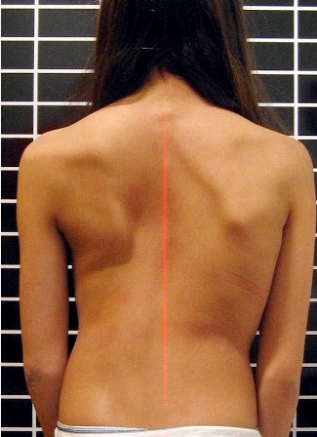
Patient with scoliosis. Note shift relative to red line.
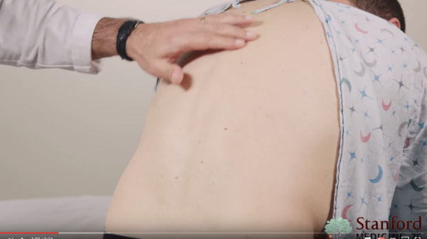
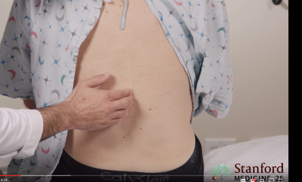
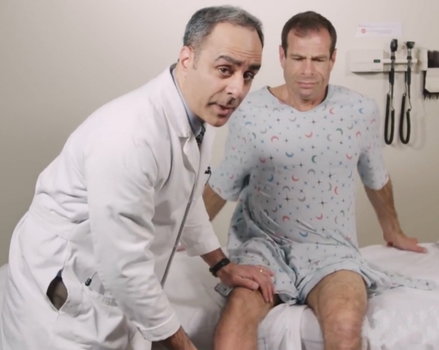
Palpation
Low Back Exam
For the second part, palpation, we generally focus on two areas:
1) The center of the back or the spinal region. Pain here suggests pain from the from the vertebra
2) Just lateral to the center or para-spinal regions. Pain here suggests pain from a muscle strain of the paraspinal muscles.
Click this link to jump to the section on inspection in the video.
Palpation of the spine.
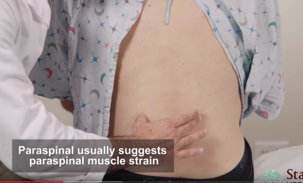
Palpation of paraspinal region.
Provocative Tests
Low Back Exam
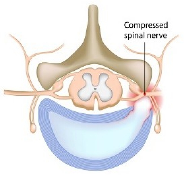
The purpose of provovative tests is to elicit pain by specific manuvers, thus a positive test. If positive, these manuvers suggest the nerve is being irritated by a mechanical cause, usually the verebral bones or herniated disc. The irritative nerves form the sciatic nerve, leading to sciatica.
Click this link to jump to the section on provocative tests in the video.
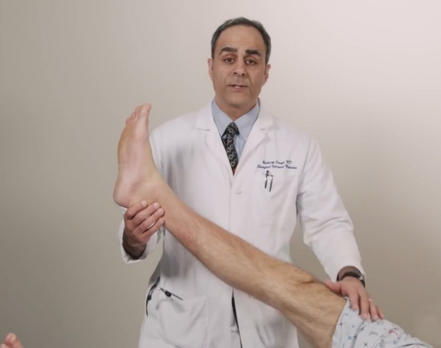
Straight Leg Test
The most common provocative test is the straight leg test. To conduct this test, have the pateint lay supine and passively elevate the fully extended leg of the affected side to 30-60 degrees. You should need to extend the leg more than 60 degrees. A positive test will elicit pain in the region where the patient was complaining of pain in the back, often radiating down the leg.
You can often elicit pain of the affected side by lifting the leg on the other side if the nerve irritation is severe enough.
Straight Leg Test Variant
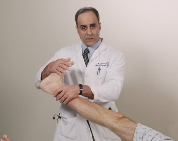
Another variant of the straight leg test involves lower the leg to around 30 degrees and flexing the foot and depicted in the image. As in the straight leg test, a positive test involves pain in the lower back, often radiating down the leg.
Tripod Sign
The tripod sign is a provocative test that is conducted while the patient is in the seated position. By elevating one of the legs, a positive sign will elicit pain in the back (again often radiating down the leg) and should be accompanied by the patient's natural tendency to decrease the pain by leaning back and resting both arms on the table to support him or herself, thus the creating a tripod.
Note: this is a good sign to use with patient's suspected of malingering if they complain of pain. Failure to lean back and rest both arms on the table may suggest the pain is note present or not related to irritation of the nerve roots.
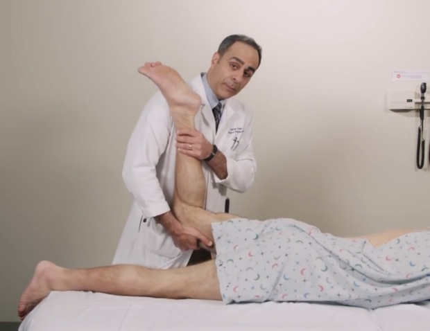
Femoral Stretch Test (L2-4)
The straight leg and tripod signs are more sensitive for pain in the L5 & S1 regions. If you suspect pain coming from the L2-4 region (which is less common), you can test for it with the femoral stretch test.
This test is done having your patient lie prone on their stomach. Next, flex the leg at the knee while holding the base of the lef under the knee. Next, simply lift the whole leg up. A positive test suggests pain in the L2-4 region if they complain of pain in the anterior thigh while the leg is lifted up. (See image. Or click this link to jump to this section on the video.)
Neurological Exam
Low Back Exam
For many patients, palpation and provocative tests are enough to confirm a musculoskeletal cause. Conservative treatment is then prescribed. However, if your patient has severe or prolonged pain or if there is any concern from the history about neurological dysfunction a neurological exam should be conducted.
The neurological exam consists of the:
1) Motor Exam
2) Sensory Exam
3) Reflex Exam
Of note, the major nerve roots to examine include L4, L5 and S1 as they are the most commonly affected. Therefore, we will focus on these three roots as well for each neurological exam.
Click this link to jump to the section on the neurological exam in the video.
Motor Exam
In Low Back Pain
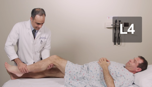
L4 Motor Exam
To test L4 strength, have the patient slightly bend the knee and kick out as you keep pressure against the leg. Be sure to compare both sides to see if one side has weakness relative to the other.
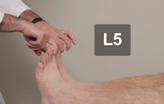
L5 Motor Exam
To test L5 strength, hold pressure over the large toes and ask the patient to dorsiflex the big toes and foot towards up. Compare both sides for relative weakness.
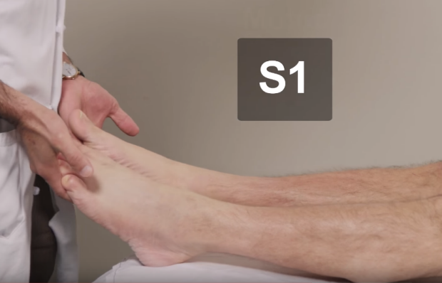
S1 Motor Exam
To test S1 strength, hold pressure under both feet and ask the patient to plantarflex the foot down. Compare both sides for relative weakness.
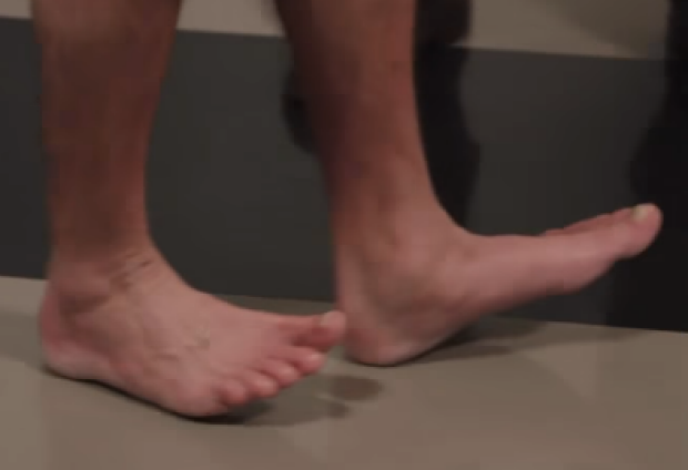
Test for L5 weakness with walking on heels in normal patient. If one foot is unable to lift toes off ground, could suggest L5 weakness on that side.
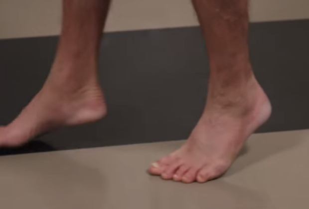
Test for S1 weakness with walking on toes in normal patient. If one foot is unable to lift heal off ground, could suggest S1 weakness on that side.
Sensory Exam
In Low Back Pain
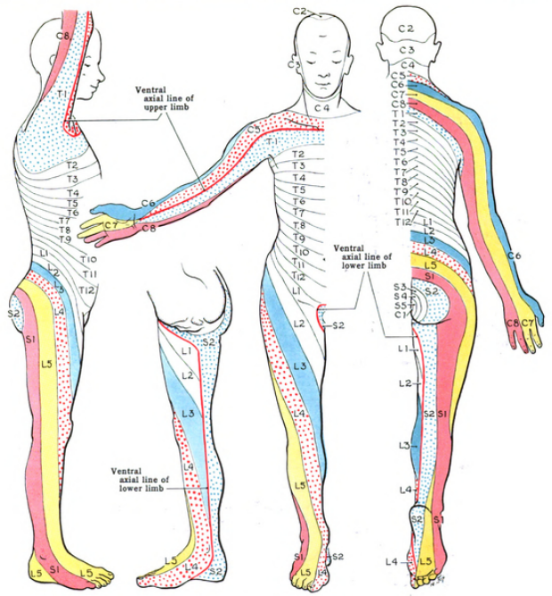
In the sensory exam, again focusing on L4, L5 & S1, we will look at specific dermatomal regions as noted in the image. If possible, use a monofilament. If not present, you can use your fingers or the tip of a tongue depressor to test for sensation.
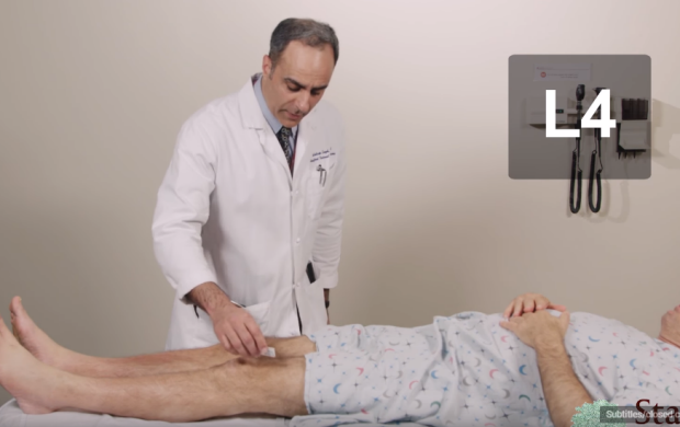
L4 Sensory Exam
Focus on the anterior/lateral aspect of the thigh.
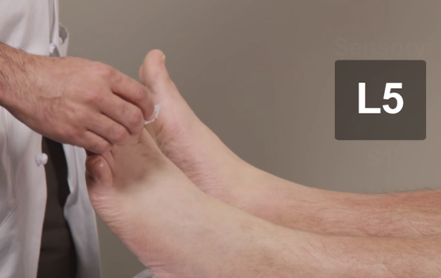
L5 Sensory Exam
Focus on the space on the dorsal side between the first and second toe.
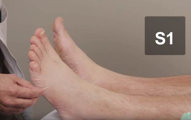
S1 Sensory Exam
Focus on the lateral aspect of the foot.
Reflex Exam
In Low Back Pain
The last part of the neurological assessment is the reflex exam. Again we look at L4, L5 & S1. Pay attention to differences on either side.
Click here to visit our page about the deep tendon reflex exam.
Click here to jump to the section on reflexes on the low back pain video.
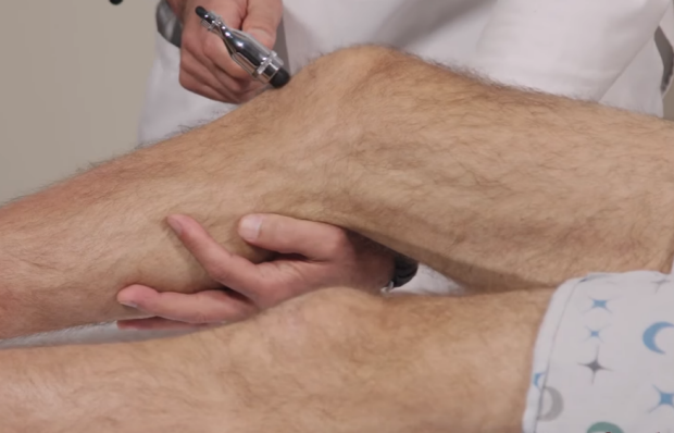
L4 Reflex Exam
L4 is tested by the patellar reflex.
L5 Reflex Exam
L5 is tested by the medial hamstring reflex.
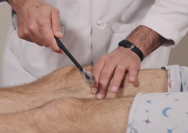
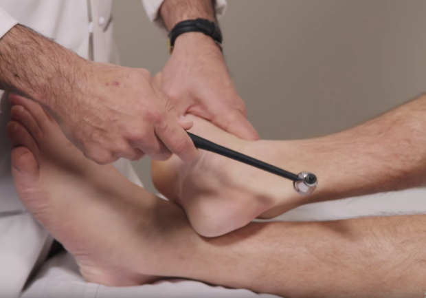
S1 Reflex Exam
L4 is tested by the achilles reflex.
Consult the Expert on Low Back Pain

Baldeep Singh
Dr. Baldeep Singh is a Clinical Professor at Stanford University and the Vice Chair for Academic Affairs for the Division of Primary Care and Population Health. He is involved in a number of clinical teaching roles at Stanford's School of Medicine and an active member of the Stanford Medicine 25 team with a special expertise in the exam of the lower back and regional hip pain.
Clinical Pearl
When a patient with low back pain has a normal neurological exam, imaging such as MRI or referral to specialists are often not necessary. Conservative treatment such as an ergometric evaluation, attention to exercise modification, physical therapy, and occasional use of NSAIDS and/or acetaminophen are often the focus of treatment.
Differential Diagnoses
Low Back Exam
While by far the most common causes of low back pain are related to the muscle or bone (that is, less worrisome causes from a diagnostic standpoint), it's important to remember the other causes of back pain that may be suggested by the history, physical exam findings or additional tests.
These diagnoses include:
- Malignancy
- Infection (such as an osteomyelitis of the lumbar spine)
- Inflammatory Arthritis
- Back Pain Mimickers:
- Prostatitis
- Pelvic Inflammatory Disease
- Kidney Stones
- Aortic Abdominal Aneurysm
- Gastrointestinal Disease
Key Learning Points
Learn the low back exam aspects that include:
- inspection
- palpation
- provocative tests
- neuro exam
- awareness of more concerning causes
Related to Low Back Exam
The Stanford Medicine 25
- Aortic Regurgitation Exam
- Ankle Brachial Index
- Ankle and Foot Exam
- Ascites & Venous Patterns
- Bedside Ultrasound
- Breast Exam
- Cardiac Second Sounds
- Carpal Tunnel Exam
- Cerebellar Exam
- Deep Tendon Reflexes
- Dermatology Exam: Acne vs. Rosacea
- Dermatology Exam: Learning the Language
- Dermatology Exam: Nevi (Mole) Exam
- Fundoscopic Exam (Ophthalmoscopy)
- Gait Abnormalities
- Hand Exam
- Hip Region Exam
- Internal Capsule Stroke
- Involuntary Movements and Tremor Diagnosis: Types, Causes, and Examples
- Knee Exam
- Liver Exam
- Low Back Exam
- Lymph Node Exam
- Neck Vein Exam
- Pelvic Exam
- Precordial Movements in the Cardiac Exam
- Pulmonary Exam: Percussion & Inspection
- Pupillary Responses
- Pulsus Paradoxus and Blood Pressure Measurement Techniques
- Rectal Exam
- Spleen Exam
- Tarsal Tunnel Exam
- Thyroid Exam
- Tongue Exam
- Liver Disease, Head to Foot
- Visit the 25
- Shoulder Exam Tutorial
- Parkinson's Disease Exam
- Diastolic Murmurs Exam
- Dermatology Exam: Nevi (Mole) Exam