Approach to the Nevi (Mole) Exam
All nevi (or moles) should be taken seriously as they may be normal or represent a cancer. Here we review the characteristics of any mole that should increase suspicion of cancer.
Introduction to the Nevi (Mole) Exam
Normal Findings with Moles
While looking for abnormal lesions, you may stumble upon these normal ones.
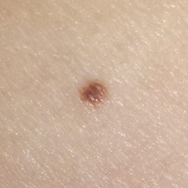
Normal mole
Symmetric, small than 6 mm, one color. These are the hallmarks of a normal mole. See below for signs of abnormal lesions.
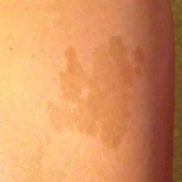
Seborrheic keratosis
These are common, benign skin lesions that occur more frequently with age. The are often described as "stuck-on" lesions, as if a ball small ball of gum was thrown again a wall and stuck to it. There is nothing needed to do for these lesions other than reassure your patient. (Note: patients with many of these, such as greater than 50, often all over torso can be a sign of a hidden malignancy, usually with the gastrointestinal tract. This is referred to as the sign of Leser-Trelat.)
Cherry hemangioma
A normal and very common lesion, these are caused by the proliferation of small blood vessels in the skin. There is no need for and medical intervention for these.
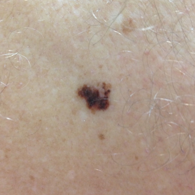
Abnormal Findings - ABCDE's of Melanoma
These signs should lead you to monitor skin lesions more closely or have them biopsied to diagnose melanoma.
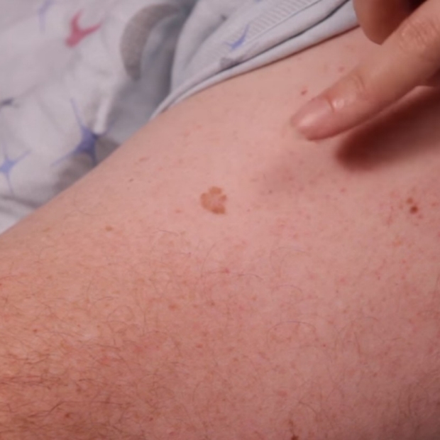
Asymmetry
If you were to cut a line down the middle of this lesion, both halves will NOT look like a mirror image of each other.
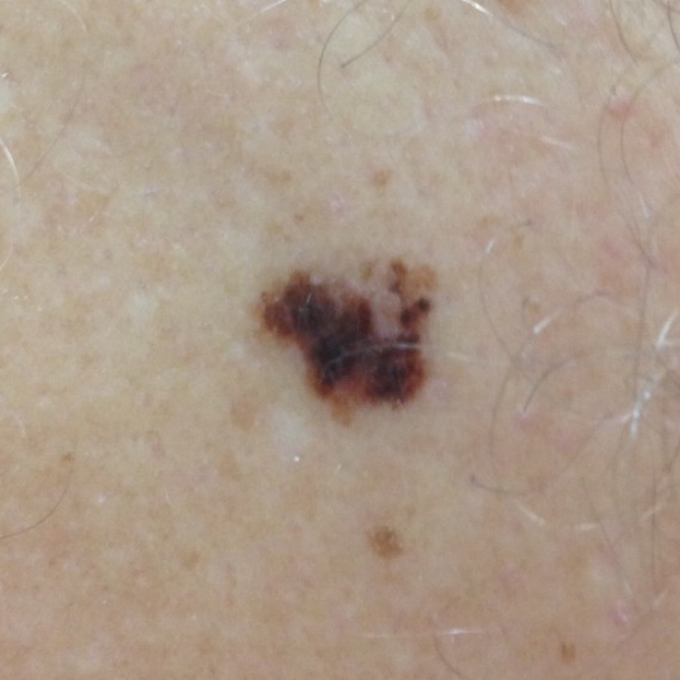
Border (irregular)
Color
Multiple colors in one lesion can suggest underlying melanoma.
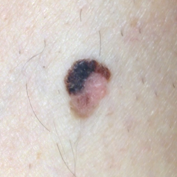
Diameter
Usually greater than 6mm, which is about the size of a pencil eraser. However, remember small moles do not rule out melanoma.
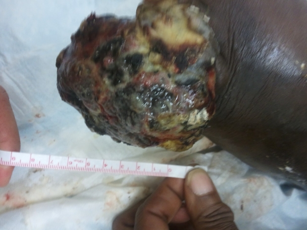
Evolving
This is the most important sign! If a lesion is changing (e.g. growing, new colors or change in border), you should keep a high suspicion for melanoma.
.gif)

The Ugly Duckling Sign
This refers to one mole among many that sticks out and looks different ("the ugly duck") and should raise suspicion for melanoma.
.gif)
Consult the Expert

Jennifer Chen
Dr. Chen is a Clinical Assistant Professor and Dermatologist at Stanford University Hospital and Clinics.
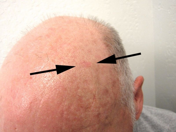
Clinical Pearl
When you examin the skin, always get your fingers involved to palpate lesions that are harder to see. A classic example of this is the skin cancer precursor called an actinic keratosis (image below). In this lesion, you often feel it before it becomes easier to see.
Key Learning Points
- Learn the general approach to the nevi (mole) exam
- Learn the signs of an abnormal lesion
Related to Dermatology Exam
The Stanford Medicine 25
- Aortic Regurgitation Exam
- Ankle Brachial Index
- Ankle and Foot Exam
- Ascites & Venous Patterns
- Bedside Ultrasound
- Breast Exam
- Cardiac Second Sounds
- Carpal Tunnel Exam
- Cerebellar Exam
- Deep Tendon Reflexes
- Dermatology Exam: Acne vs. Rosacea
- Dermatology Exam: Learning the Language
- Dermatology Exam: Nevi (Mole) Exam
- Fundoscopic Exam (Ophthalmoscopy)
- Gait Abnormalities
- Hand Exam
- Hip Region Exam
- Internal Capsule Stroke
- Involuntary Movements and Tremor Diagnosis: Types, Causes, and Examples
- Knee Exam
- Liver Exam
- Low Back Exam
- Lymph Node Exam
- Neck Vein Exam
- Pelvic Exam
- Precordial Movements in the Cardiac Exam
- Pulmonary Exam: Percussion & Inspection
- Pupillary Responses
- Pulsus Paradoxus and Blood Pressure Measurement Techniques
- Rectal Exam
- Spleen Exam
- Tarsal Tunnel Exam
- Thyroid Exam
- Tongue Exam
- Liver Disease, Head to Foot
- Visit the 25
- Shoulder Exam Tutorial
- Parkinson's Disease Exam
- Diastolic Murmurs Exam
- Dermatology Exam: Nevi (Mole) Exam