Approach to the Hip Region Exam
Hip pain is really common. Sometimes the pain is coming from the hip joint. Other times, pain may be coming from other surrounding causes. Here, we'll review all causes of pain in the hip region and show you the exam to diagnose each one.
Introduction
Pain in the Hip Region
Before we can review the exam of the hip, it's important to know the possible diagnoses. That way you're going through each exam maneuver unique to each specific diagnosis. To aid in the process, it's helpful to organize your diagnoses into three locations: anterior, lateral and posterior.
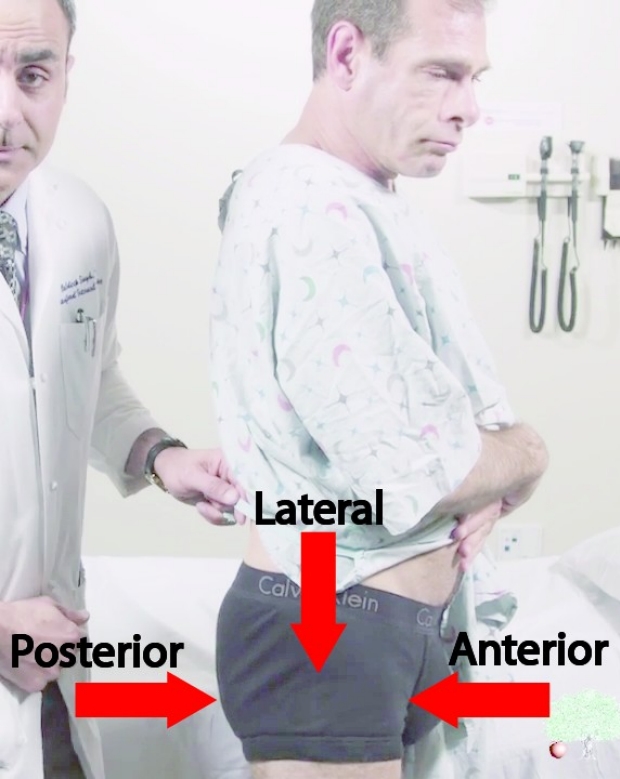
Differential Diagnoses of Regional Hip Pain
Anterior
- Hip Joint Disease, including:
- Osteoarthritis
- Neck Fracture
- Septic Arthritis
- Femoral-Acetabular Syndrome
- Avascular Necrosis
- Meralgia Paresthetica (can be anterior and/or lateral)
Lateral
- Greater Trochanteric Bursitis
- External Snapping Hip
Posterior
- Radiculopathy (visit Low Back Exam page)
- Sacroiliitis or Sacraliliac Joint Pain
- Piriformis Syndrome
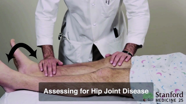
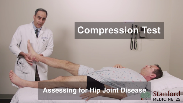
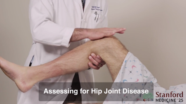
Hip Joint Disease
Anterior Region
- Pain coming from the hip joint usually presents in the anterior region. As noted above, there are a number of problems that could arise in the hip joint (e.g. osteoarthritis, neck fracture, septic arthritis femoral-acetabular syndrome and avascular necrosis). Together with the exam noted below, once you are able to localize the pain to the hip joint, you can use other data (e.g. vitals, labs and imaging) to help with the specific diagnosis.
- Below are the number of exam manuevers used to localize pain to the hip joint.
- Click this link to jump to the section on anterior hip pain in the video.
Log Rolling Test
The log rolling test can be helpful if you suspect a hip fracture. It is done by simply rolling the leg side-to-side while the patient is supine.
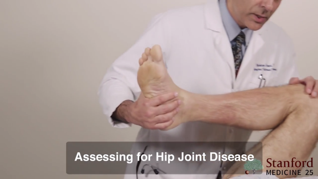
Compression Test at the Foot
The compression test is done by hitting the balls of the feet to transmit pressure towards the hip while the leg is lifted. Again, this can be very painful in a hip fracture.
Compression Test at the Knee (and Lateral Hip)
The compression test can also be done by tapping at the knee, again transmitting pressure towards the hip. Finally, you can also by tap lateral to the hip (see video for demonstration). All compression tests can be positive in hip pain but especially with a fracture. If positive, hip x-rays (and possibly more imaging) should be ordered.
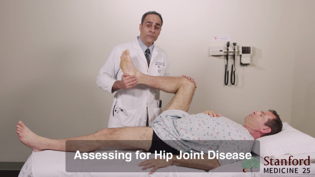
Range of Motion of Hip Joint
Video demonstration to test range of motion.
Tests for range of motion look for pain in the hip joint as the ball-and-socket joint of the hip is activated. The most common cause of pain with range of motion is osteoarthritis (or degenerative joint disease) of the hip but again any of the problems of the hip listed above will likey cause pain during range of motion and/or limitation in range of movement.
Some of these motions include:
- Flexing both hip and knee at 90 degress, then rotating back-and-forth both interally and externally to approx 45 degrees.
- Flexing the hip and knee beyond 90 degress to the extremes of range of motion and rotating from side-to-side
Range of motion at 90 degrees hip and knee flexion.
Extreme range of motion beyond 90 degrees hip and knee flexion.
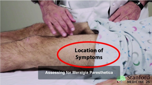
Meralgia Paresthetica
Anterior/Lateral Region
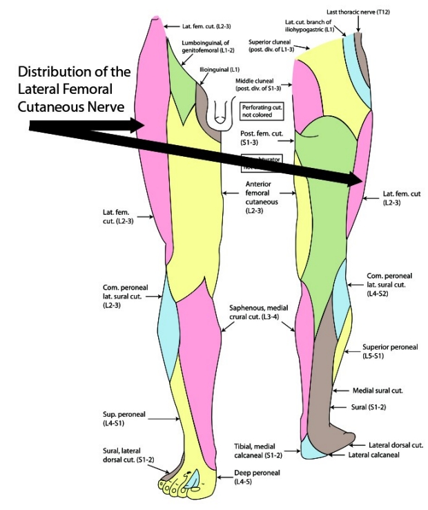
If your patient complains of anterior (sometimes anterior and lateral) thigh pain, the diagnoses may be meralgia paresthetica. This diagnosis is usually caused by entrapment of the lateral femoral cutaneous nerve as it passes under the inguinal ligament and supplies the sensory distribution of the anterior/lateral thigh.
The pain is often described as burning. There may be an associated paresthesia (numbness and/or tingling) and sometimes a decrease in sensation. Therefore, a sensory exam should be performed in the region. Meralgia paresthetica is more common in diabetics.
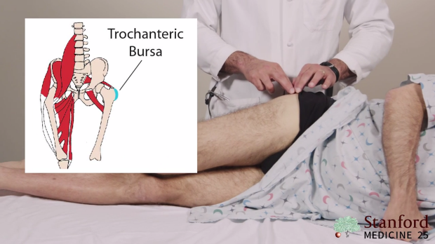
Greater Trochanteric Bursitis
Lateral Region
Greater trochanteric bursitis consists of pain over the lateral side of the hip. The diagnosis of greater trochanteric bursitis is diagnosed by asking the patient to lay on the side (painful side up) and palpating over the burse of the greater trochanter. Tenderness should be appreciated. Pain is often self-limiting but can also be treated with NSAIDs.
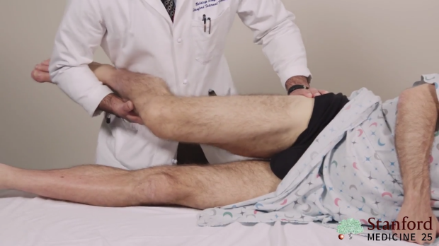
External Snapping Hip
Lateral Region
Greater trochanteric bursitis has more recently been thought to be part of a greater trochanteric pain syndrome that includes the tendons that insert and cross over the greater trochanter such as the iliotibial band which traverses the lateral hip and if inflammed could cause a "snap" during movement as the hip flexes and extends.
To test for an external stapping hip, with the patient on their side (painful side up), you will grab the whole leg then flex and extend the leg while palpating the iliotibal tendon (near the greater trochanter) and feeling for a popping or snapping that may be associated with pain. Link to video demonstrating this.
Lumber Radiculopathy
Posterior Region
A very common cause of posterior pain near the buttocks is caused by radiculopathy as the nerves exit the lumber spine. That topic is covered in our page on the Approach to Low Back Pain here.
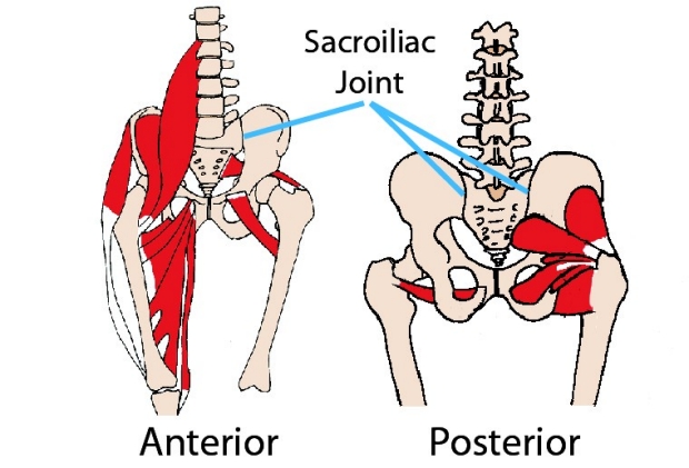
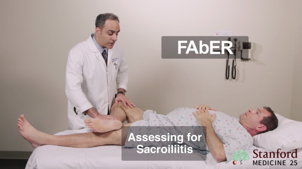
Sacroiliitis / Sacraliliac Joint Pain
Posterior Region
Pain at the sacroiliac joint is often in the posterior or buttocks region. Pain in the sacroiliac joint can be elicited with the FAbER Test. FAbER stands for Flexion, Abduction and External Rotation. Once the leg is flexed, abducted and externally rotated (as noted in image below), you will apply a downward pressure at the knee. The presence of pain in the posterior (or buttocks) region suggests pain from the sacroiliac joint.
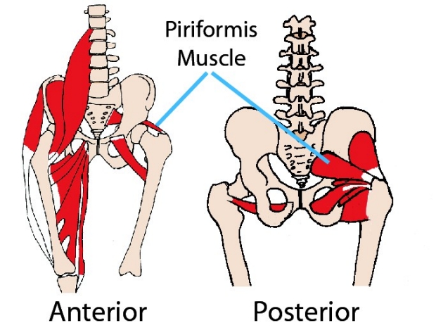
Piriformis Syndrome
Posterior Region
Piriformis syndrome occurs when the piriformis muscle compresses the sciatic nerve. Tests for piriformis sydrome are attempting to move the leg so that the piriformis muscle pushes against the sciatic nerve leading to pain and thus a positive test.
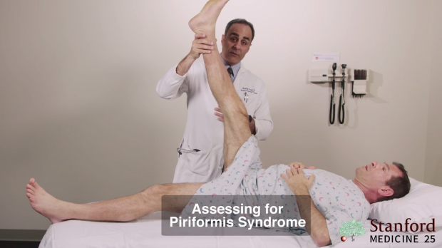
In the first manuver, keeping the leg straight, flex the hip up to 90 degrees, looking for pain in the posterior/buttocks region.
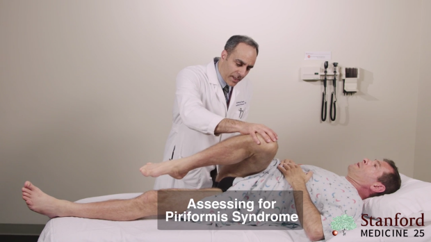
In the second manuver, keeping the hip flexed, flex the knee and adduct the knee accross the body of the patient, again looking for pain in the the posterior/buttocks region.
Consult the Expert on Regional Hip Pain

Baldeep Singh
Dr. Baldeep Singh is a Clinical Professor at Stanford University and the Vice Chair for Academic Affairs for the Division of Primary Care and Population Health. He is involved in a number of clinical teaching roles at Stanford's School of Medicine and an active member of the Stanford Medicine 25 team with a special expertise in the exam of the lower back and regional hip pain.
Clinical Pearl
As you see from this page, most of what we refer to as "hip pain", doesn't involve the hip joint. Remember, if the pain is lateral or posterior (near buttocks), look for the other causes of pain that don't involve the hip joint.
Key Learning Points
Learn the approach to regional hip pain that includes:
- Knowing the differential diagnoses
- Knowing each unique exam that will confirm or exclude each diagnosis
Related to Low Back Exam
The Stanford Medicine 25
- Aortic Regurgitation Exam
- Ankle Brachial Index
- Ankle and Foot Exam
- Ascites & Venous Patterns
- Bedside Ultrasound
- Breast Exam
- Cardiac Second Sounds
- Carpal Tunnel Exam
- Cerebellar Exam
- Deep Tendon Reflexes
- Dermatology Exam: Acne vs. Rosacea
- Dermatology Exam: Learning the Language
- Dermatology Exam: Nevi (Mole) Exam
- Fundoscopic Exam (Ophthalmoscopy)
- Gait Abnormalities
- Hand Exam
- Hip Region Exam
- Internal Capsule Stroke
- Involuntary Movements and Tremor Diagnosis: Types, Causes, and Examples
- Knee Exam
- Liver Exam
- Low Back Exam
- Lymph Node Exam
- Neck Vein Exam
- Pelvic Exam
- Precordial Movements in the Cardiac Exam
- Pulmonary Exam: Percussion & Inspection
- Pupillary Responses
- Pulsus Paradoxus and Blood Pressure Measurement Techniques
- Rectal Exam
- Spleen Exam
- Tarsal Tunnel Exam
- Thyroid Exam
- Tongue Exam
- Liver Disease, Head to Foot
- Visit the 25
- Shoulder Exam Tutorial
- Parkinson's Disease Exam
- Diastolic Murmurs Exam
- Dermatology Exam: Nevi (Mole) Exam