Cardiac Second Heart Sounds
The cardiac second sounds can provide a number of valuable clues to what is going on with the heart. Diagnoses like pulmonary hypertension, severe aortic stenosis, an atrial septal defect and delays in the electrical conduction can be diagnosed or suspected with close attention to second heart sounds.
Introduction to Second Heart Sounds
The cardiac second sound (the "dub") can be a source of some confusion. However, the subtle changes of the second heart sound can tell you a great deal about the heart. Here we discuss the basics of listening for the second heart sounds and review abnormal findings.
The Basics of Second Heart Sounds
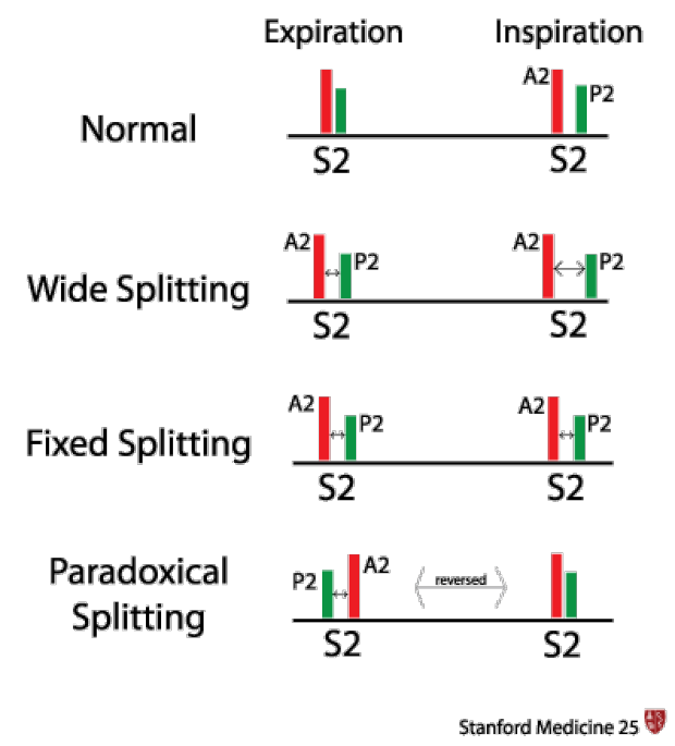
In the normal heart:
During expiration:
The second sound (S2) is usually single
During inspiration:
The second sound (S2) is made of two component sounds:
Aortic valve closure (A2) which happens first.
Pulmonic valve closure (P2) which happens second.
A2 is heard widely all over the chest. So when you hear 'S2' at the mitral area, you are really hearing A2.
Normally, P2 is soft and only heard at the pulmonic region (left parasternal, intercostal space 2), however even in this region A2 is louder.
.gif)
What causes the split second heart sounds?
There are believed to be multiple causes for the physiologic splitting of S2. Both A2 and P2 close when the pressure above the respective valves are greater than the pressure in the ventricles below. Given the lower vascular resistance of the pulmonary artery, during inspiration, the pulmonary artery is able to tolerate more volume of blood before the pressure above the valve increases. Additionally, during inspiration, more blood fills the right ventricle leading to a slightly longer ejection time, adding to the delayed pulmonic valve closure.
.gif)
Exam Technique in Second Heart Sounds
- Splitting best heard in the 2nd left intercostal space, close to the sternal border.
- Use the diaphragm of your stethoscope
- Second heart sounds are best heard when patients are semi-recumbent (30-40 degrees upright) and in quiet inspiration.
- The intensity of P2 is determined relative the A2. The intensity of P2 is considered elevated if P2 is louder than A2 at the pulmonic region (left parasternal, intercostal space 2)
Pathological Findings in Second Heart Sounds
- Increased intensity of P2: P2 louder than A2 at pulmonic region (left parasternal, intercostal space 2)
- DDx: pulmonary hypertension (most common), ASD will also increase P2
- Note: Since P2 is measured relative to A2, causes for lower A2 intensity should be ruled out. These include: mitral regurgitation, aortic regurgitation, low diastolic arterial pressure, severe immobile aortic valve disease
- DDx: pulmonary hypertension (most common), ASD will also increase P2
- Wide splitting: Detected by presence of splitting during expiration, wider during inspiration
- DDx: Anything that causes delayed conduction down the right bundle (RBBB, pre-excitation of left ventricle, pacing of left ventricle, premature LV beats), pulmonary stenosis, pulmonary arterial hypertension
- Fixed splitting: Spitting at both expiratory and inspiratory phases but does NOT lengthen with inspiration
- Dx: ASD (due to continuous blood flow from left side to right side leading lenthened cardiac cycle on the right side of the heart), Right heart failure, Pulmonary Hypertension
- Paradoxical splitting: Reverse of normal physiology, splitting of second heart sounds during expiration, singular during inspiration
- Dx: Anything that causes delayed conduction down the left bundle (LBBB, pre-excitation of right ventricle, right ventricular pacing, premature RV beats), aortic stenosis
- Single S2: Either from loss of A2 or loss of P2
- DDx: Severe aortic stenosis, severe aortic regurgitation, congenital absence of pulmonary valve
- Note: in patients with difficult to hear heart sounds (obesity, emphysema, pericardial fluid), P2 may be too hard to hear causing a single (A2) heart sound
- DDx: Severe aortic stenosis, severe aortic regurgitation, congenital absence of pulmonary valve
Key Learning Points
- Basic physiology and technique for listening to second heart sounds
- Key findings seen in the abnormal second heart sounds
Related to Cardiac Second Sounds
The Stanford Medicine 25
- Aortic Regurgitation Exam
- Ankle Brachial Index
- Ankle and Foot Exam
- Ascites & Venous Patterns
- Bedside Ultrasound
- Breast Exam
- Cardiac Second Sounds
- Carpal Tunnel Exam
- Cerebellar Exam
- Deep Tendon Reflexes
- Dermatology Exam: Acne vs. Rosacea
- Dermatology Exam: Learning the Language
- Dermatology Exam: Nevi (Mole) Exam
- Fundoscopic Exam (Ophthalmoscopy)
- Gait Abnormalities
- Hand Exam
- Hip Region Exam
- Internal Capsule Stroke
- Involuntary Movements and Tremor Diagnosis: Types, Causes, and Examples
- Knee Exam
- Liver Exam
- Low Back Exam
- Lymph Node Exam
- Neck Vein Exam
- Pelvic Exam
- Precordial Movements in the Cardiac Exam
- Pulmonary Exam: Percussion & Inspection
- Pupillary Responses
- Pulsus Paradoxus and Blood Pressure Measurement Techniques
- Rectal Exam
- Spleen Exam
- Tarsal Tunnel Exam
- Thyroid Exam
- Tongue Exam
- Liver Disease, Head to Foot
- Visit the 25
- Shoulder Exam Tutorial
- Parkinson's Disease Exam
- Diastolic Murmurs Exam
- Dermatology Exam: Nevi (Mole) Exam