The General Dermatology Exam: Learning the Language
The diagnosis of any skin lesion starts with an accurate description of it. To do that, you need to know how to describe a lesion with the associated language. This language, reviewed here, can be used to describe any skin finding.
Approach to General Exam
Approach to Multiple Rashes
Introduction to the Dermatology Exam
Before you can make a diagnosis of any skin lesion, it's important to be able to accurately describe the skin lesion. The purpose of this page is to cover the fundamental knowledge you will need to do this.
Learn the Language of the Dermatology Exam
- Primary Morphology
- Macule - flat lesion less than 1 cm, without elevation or depression
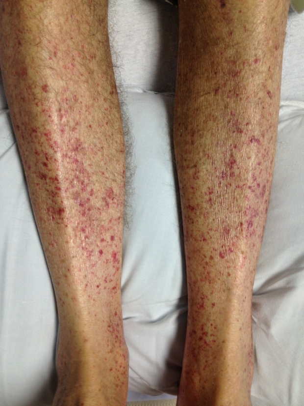
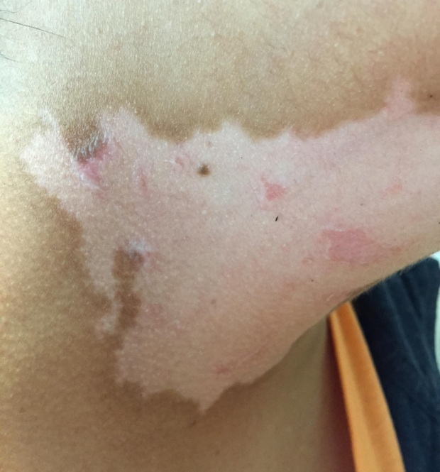
- Patch - flat lesion greater than 1 cm, without elevation or depression
- Plaque - flat, elevated lesion, usually greater than 1 cm
- Papule - elevated, solid lesion less than 1 cm
- Nodule - elevated, solid lesion greater than 1 cm
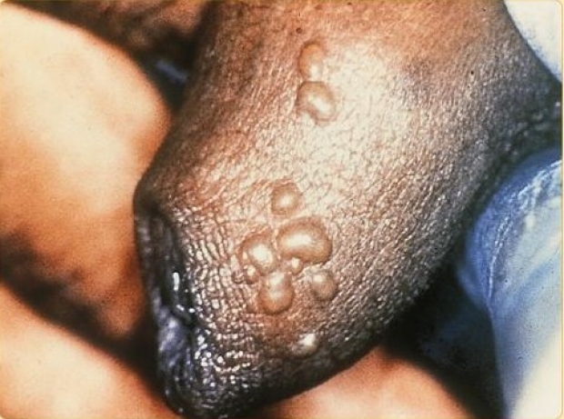
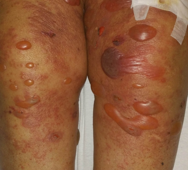
- Vesicle - elevated, fluid-filled lesion, usually less than 1 cm
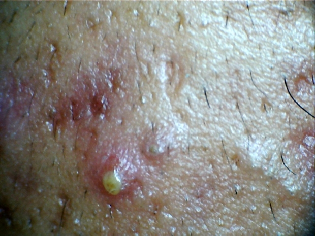
- Pustule - elevated, pus-filled lesion, usually less than 1 cm
- Bulla - elevated, fluid-filled lesion, usually greater than 1 cm
- Size
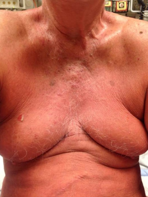
- Demarcation
- Well-demarcated
- Not well-demarcated
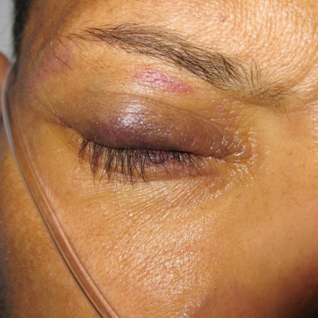
- Color
- Secondary Morphology
- Distribution
Primary Morphology

Size
How Small?
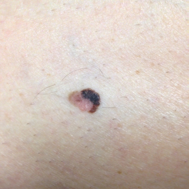
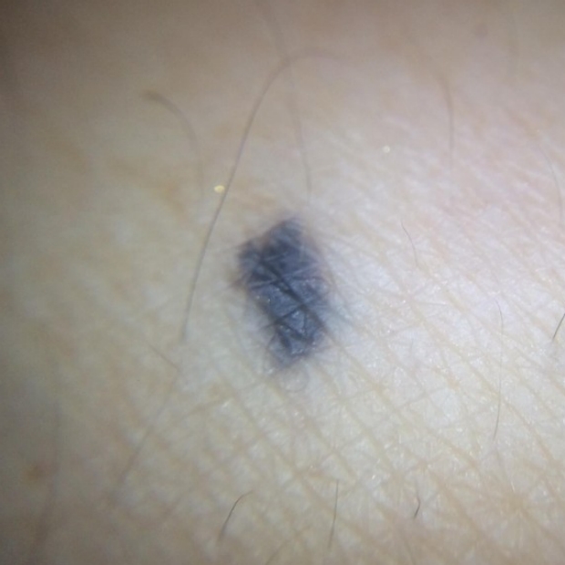
Example: Superficial Spreading Melanoma
How Large?
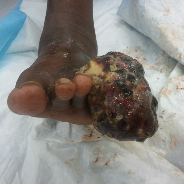
Example: Acral Lentiginous Melanoma
Demarcation
Clearly Defined?
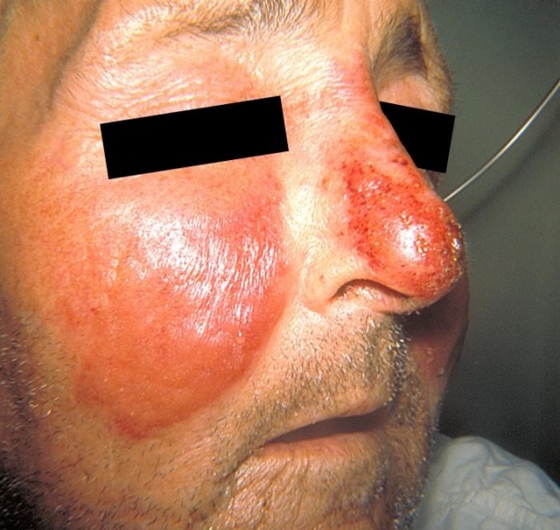
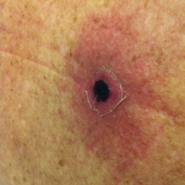
Example: Erysipelas
Not Well Defined?
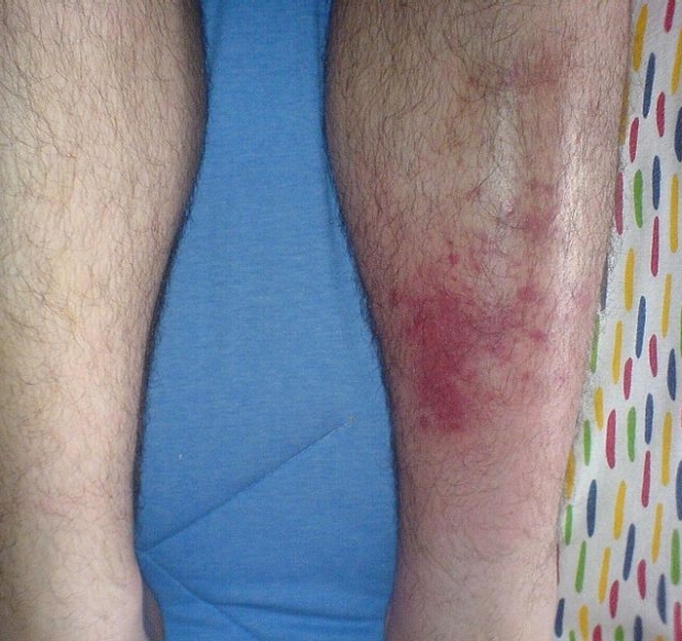
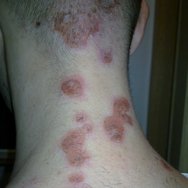
Example: Cellulitis
Consult the Expert

Justin Ko
Dr. Justin Ko is a Clinical Assistant Professor, Dermatologist and Clinic Chief and Director of Medical Dermatology at Stanford.
Clinical Pearl
A good dermatology exam requires a well lit room and sometimes a hand held light. Sometimes, you may need a ruler and magnifiying glass. Remember that sometimes the palpation of skin lesions can be just as important as what you see.
Color
Secondary Morphology
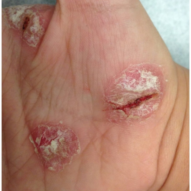

Erosion
Partial loss of epidermis.
Example: Scratching, minor skin injury

.gif)
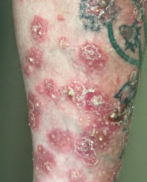
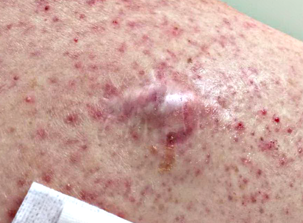
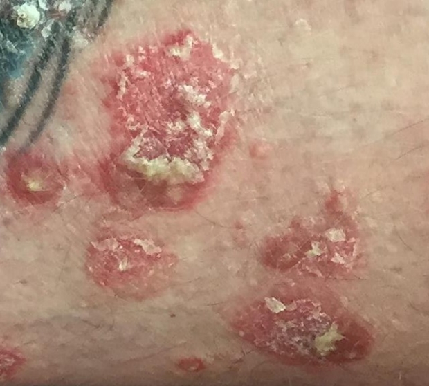
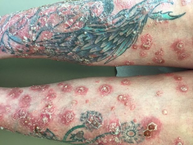
Applying What We Learned
Based on the above image, here's how we'd describe this skin lesion:
- Primary morphology -> plaque,
- Size -> a few centimeters
- Well-Demarcated
- Color -> Dully red
- Secondary morphology -> Dry serum/crusting, erosions and scaling
- Distribution -> Extensor surface of leg
Diagnosis is consistent with psoriasis given the above description.
.gif)
Key Learning Points
- Learn the language of the skin exam
- Learn to apply this approach for any skin exam
Related to Dermatology Exam
The Stanford Medicine 25
- Aortic Regurgitation Exam
- Ankle Brachial Index
- Ankle and Foot Exam
- Ascites & Venous Patterns
- Bedside Ultrasound
- Breast Exam
- Cardiac Second Sounds
- Carpal Tunnel Exam
- Cerebellar Exam
- Deep Tendon Reflexes
- Dermatology Exam: Acne vs. Rosacea
- Dermatology Exam: Learning the Language
- Dermatology Exam: Nevi (Mole) Exam
- Fundoscopic Exam (Ophthalmoscopy)
- Gait Abnormalities
- Hand Exam
- Hip Region Exam
- Internal Capsule Stroke
- Involuntary Movements and Tremor Diagnosis: Types, Causes, and Examples
- Knee Exam
- Liver Exam
- Low Back Exam
- Lymph Node Exam
- Neck Vein Exam
- Pelvic Exam
- Precordial Movements in the Cardiac Exam
- Pulmonary Exam: Percussion & Inspection
- Pupillary Responses
- Pulsus Paradoxus and Blood Pressure Measurement Techniques
- Rectal Exam
- Spleen Exam
- Tarsal Tunnel Exam
- Thyroid Exam
- Tongue Exam
- Liver Disease, Head to Foot
- Visit the 25
- Shoulder Exam Tutorial
- Parkinson's Disease Exam
- Diastolic Murmurs Exam
- Dermatology Exam: Nevi (Mole) Exam