Diagnose the cause of sepsis from one cell in the blood…
March 9, 2016
Okay so we’re usually focusing on the physical exam but in this case, we’re demonstrating how taking the time to look at a patient’s blood under a microscope can help you diagnose and treat a sick patient. In the end, it’s all about good care for your patient right?
The case:
A 45 year old woman is admitted for fever, headaches and myalgias. Her white count is elevated and she is septic. What’s the likely pathogen? How can these cells help you?

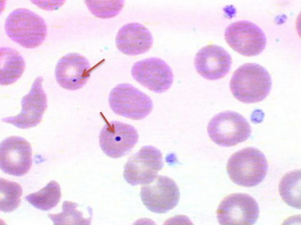
Howell-Jolly body (arrow) with red blood cells.
Answer:
This is a picture of a red blood cell with a Howell-Jolly body (red arrows). They are left over nuclear remnants that are usually removed when blood cells are in the spleen. Howell-Jolly bodies occur where there is no spleen or an non-functioning spleen, referred to as asplenia. They are usually one of these at most in a red cell, round, dark purple to red in color and often located peripherally on the red blood cell.
If a patient comes in with sepsis, fever, headache and myalgias, meningitis is very likely. The most common pathogen of meningitis is Streptococcus pneumoniae(pneumococcus). It’s also the most common infection in patients with asplenia (2). Based on the patient’s symptoms/exam and her blood smear, you have a likely diagnosis!
What are the other infections you need to consider with asplenia?
This patient is at risk for any bacteria that are encapsulated. In addition toStreptococcus pneumoniae, this includes Haemophilus influenzae and Neisseria meningitidis. Of course, the above doesn’t preclude you from empirically covering all organisms until you can confirm a diagnosis. Babesiosis is thought be more common in patients with a non-functioning spleen (3).
How is a Howell-Jolly body helpful?
Well, if you already knew she had meningitis you would already be giving the right antibiotics to cover for Streptococcus pneumoniae, Haemophilus influenzae andNeisseria meningitidis. However, the diagnosis of bacterial meningitis can often be delayed from variations in presentation. Additionally, in patients presenting with sepsis from other infections, knowing the spleen is missing can be valuable to making sure you are treating for these organisms until cultures become positive or in case you are unable to confirm the bug.
What are some causes of impaired splenic function?
This can be from either having a spleen or it not functioning:
- Congenital (rare)
- Surgical removal for trauma and diseases like (hemolytic anemia, idiopathic thrombocytopenia purpura, malignancy)
- Sickle cell disease (from auto infarction during sickle cell crises)
- Ischemic disease to spleen or infarction
- Bone marrow transplant
- Collagen vascular diseases (rare)
What are some other common red blood cells findings to look for in the blood smear?
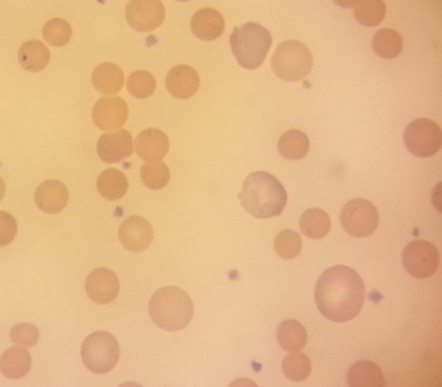
Spherocytes, noted by the lack of a pale center can been seen most commonly in hemolytic anemia (and less commonly in hereditary spherocytosis). Larger, more purple reticulocytes (see in image) will often be seen in hemolytic anemia as the body tries to make more red blood cells.
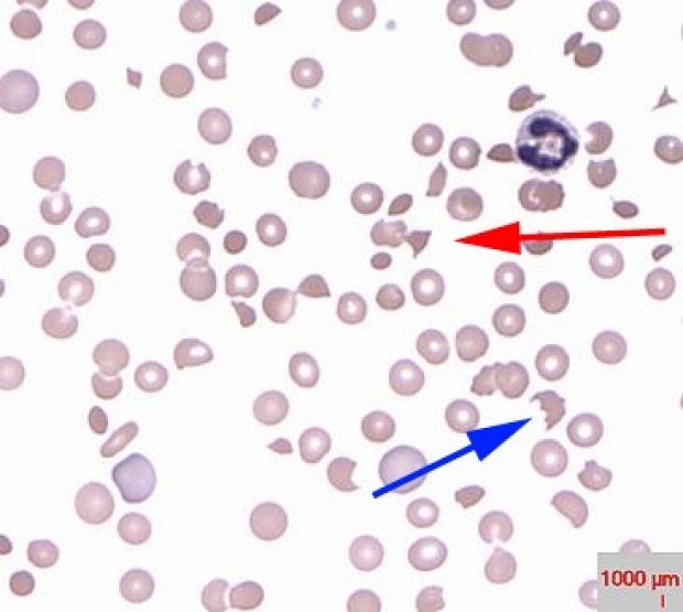
Schistocytes (red arrow) and helmet cells (blue arrow) are common in any disease where there is shearing or mechanical destruction of the red cells. This includes, disseminated intravascular coagulation, thrombotic thrombocytopenic purpura and aortic stenosis. (5)
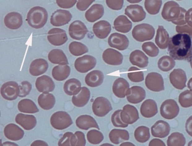
Target cells (AKA codocytes) appear as bullseyes, as seen here. These can be seen in liver disease, alpha/beta thalassemia, hemoglobin C disease and in asplenia. (6)
What is the history of Howell-Jolly?
William Henry Howell was born in Baltimore in 1860 and received a PhD from Johns Hopkins in 1884. His thesis was on the origin of fibrin in blood coagulation. During his work on coagulation, Howell began to observe how granules stained like nuclei. He went on to describe larger granules that would later be described as “Howell’s nuclear particles”. (7)
Justin Marie Jolly was born in 1870 in Melun, now a Paris suburb, obtaining his doctorate in 1898 at the University of Paris. During his work, Jolly refined and detailed many of Howell’s descriptions. He described fragmentation of nuclei during expulsion which left behind “pieces” of nuclei, and he additionally noted smaller basophilic granules.
The first report connecting the presence of a Howell-Jolly body to splenectomy was by Schur in 1908. The patient in this report suffered from pernicious anemia, hyperthyroidism and whose spleen on autopsy was found to be replaced by fibrous tissue.
What happened to the patient?
She got a lumbar puncture which had an elevated white cell count, consistent with bacterial meningitis with a negative gram stain. After getting hemodynamic support and proper antibiotics she recovered from this infection. It was thought she had a congenital version of asplenia. She was vaccinated for Streptococcus pneumoniae, Haemophilus influenzae and Neisseria meningitidis prior to discharge.
References:
- This case was obtained from Clinical Case Reports article Howell-Jolly bodies on peripheral smear leading to the diagnosis of congenital hyposplenism in a patient with septic shock
- Holdsworth RJ, Irving AD, Cuschieri A. Postsplenectomy sepsis and its mortality rate: actual versus perceived risks. Br J Surg. 1991;78(9):1031.
Rosner F, Zarrabi MH, Benach JL, Habicht GS. Babesiosis in splenectomized adults. Review of 22 reported cases. Am J Med. 1984;76(4):696.
- https://en.wikipedia.org/wiki/Howell%E2%80%93Jolly_body
- https://en.wikipedia.org/wiki/Schistocyte#/media/File:Thrombi_in_patient_with_thrombotic_thrombocytopenic_purpura_.jpg
- https://commons.wikimedia.org/wiki/File:Target_cells_and_spherocytes.jpg
- Sears, MD, Udden, MD. Howell-Jolly Bodies: A Brief Historical Review. The American Journal of the Medical Sciences Volume 343, Issue 5, May 2012, Pages 407–409