The Pelvic Exam
Pelvic exam is a important part of the exam for female patients and important towards making various diagnoses such as yeast vulvovaginitis, bacterial vaginosis, lichen sclerosis, cancers such as cervical cancer, anal/rectal cancer, fibroids, sexually-transmitted infections (gonorrhea, chlamydia, trichomonas, syphilis, herpes and human papillomavirus/genital warts) and many other diagnoses.
Introduction
Pelvic Exam
The pelvic exam is a vital part of every woman's preventative care and is also important towards making a number of diagnoses when presenting with abdominal or pelvic complaints. Here we cover each aspect of the pelvic exam and demonstrate both in text and in our video how this done.
Speculums
The main equipment you will use is the speculum so it's important for you to understand the most commonly used types. These include the:
- Pederson’s speculum
- Graves’ speculum (or Duckbill speculum) - a little wider (often used for procedure)
- Note: These are cold! Important to keep in warmer temperatures or run under warm water prior to use.
- Speculums come in various sizes, including smaller, pediatric sizes.
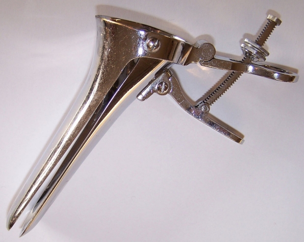
Example of a Grave's (or Duckbill) speculum which is a little wider, often used for prodecures. Image credit
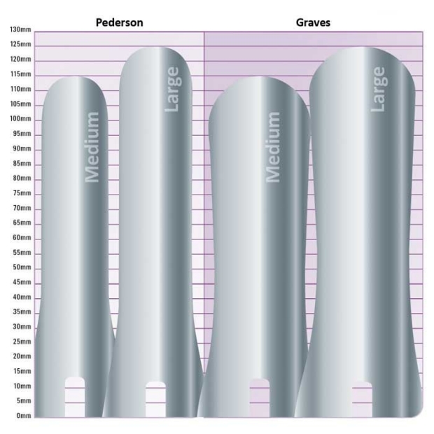
Pederson (most common) vs the Grave's (or Duckbill) speculum. Image credit
Before the Exam
Pelvic Exam
Before the exam, ensure you have:
- Speculum and lubricant
- Light source
- Needed materials if doing wet mount, pap smear or STD testing
- Have patient empty their bladder
Click this link to jump to the pre-exam section of the video.
Palpation
Pelvic Exam
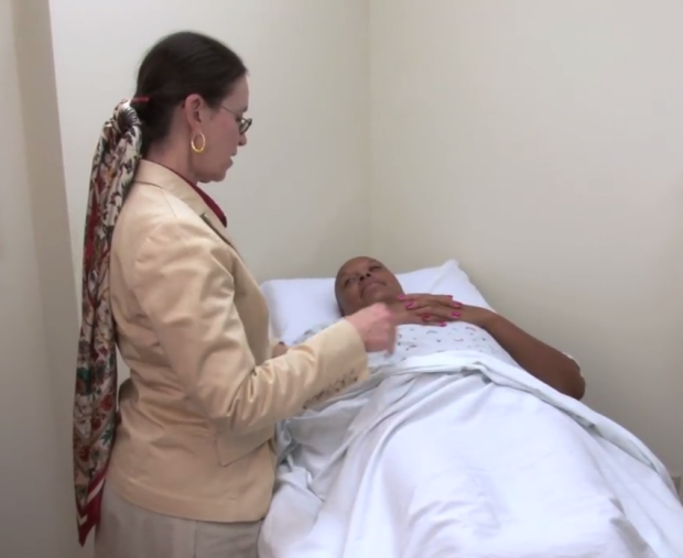
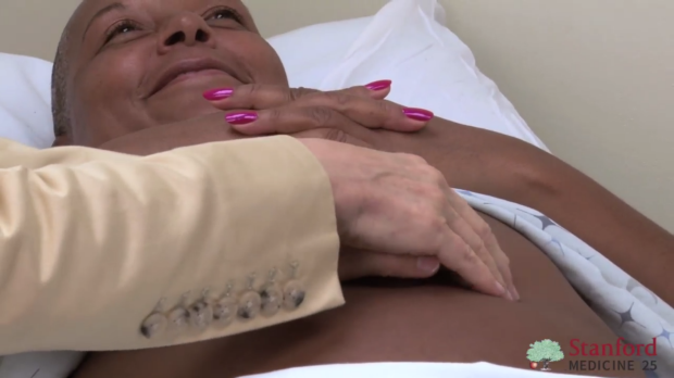
When palpating, be sure to:
o Feel for abdominal masses
o Appreciate the liver size
o Look for inguinal region for lymphadenopathy--click here for lymph node exam
Click this link to jump to the palpation section of the video.
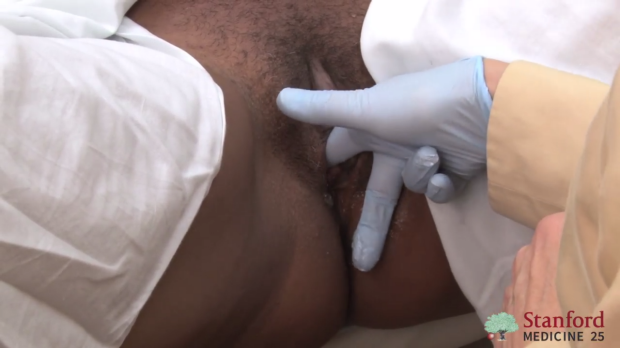
Vulvar and Speculum Examination
Pelvic Exam
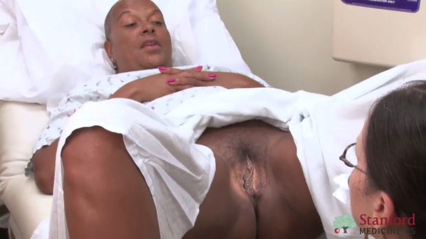
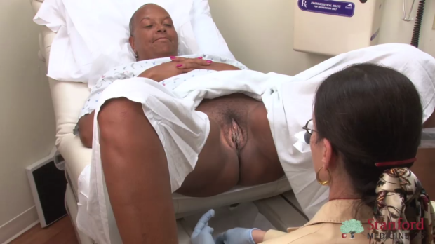
1. Examine the vulvar region looking for erythema or other rashes
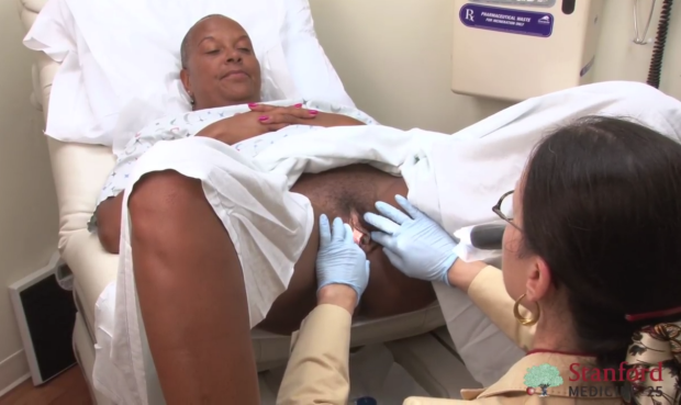
2. Place lubricant on both index finder of non-dominant hand and warmed speculum
3. Insert index finger into base of vagina
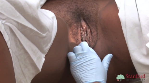
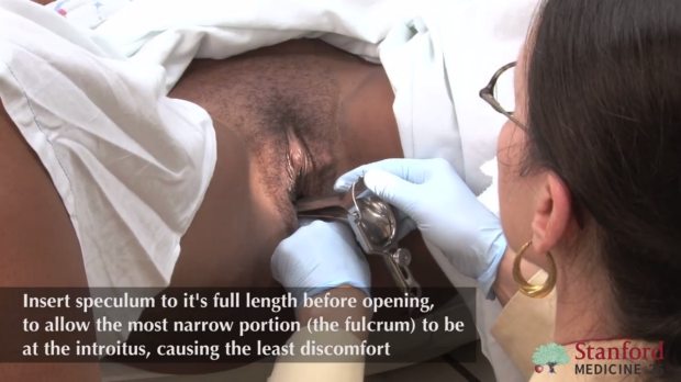
4. Touch speculum onto patient’s thigh to see if temperature appropriate then insert speculum at slight angle into vagina above finger and rotate to level position as it's inserted
Important: Insert speculum all the way BEFORE opening
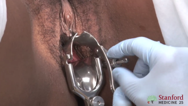
5. Open speculum and adjust until the cervix is in sight and lock open
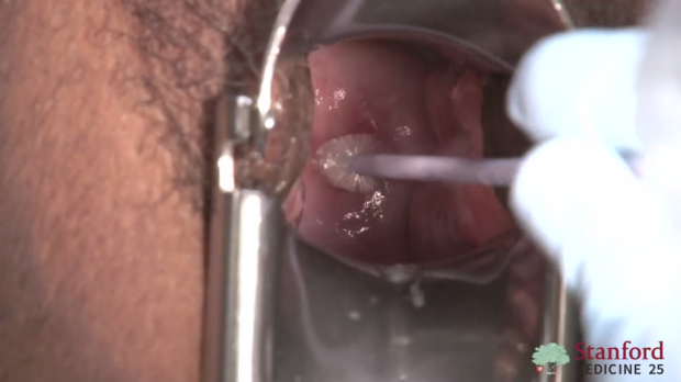
6. If planned, conduct pap smear and STD testing
Note: a little bleeding (especially if patient nulliparous) after a pap smear is expected. If bleeding profuse of cervix is erythematous and/or purulent discharge present, suspect cervicitis (gonorrhea, chlamydia).
Abnormal Findings
- Lichen Sclerosis – itching without any discharge, all age groups
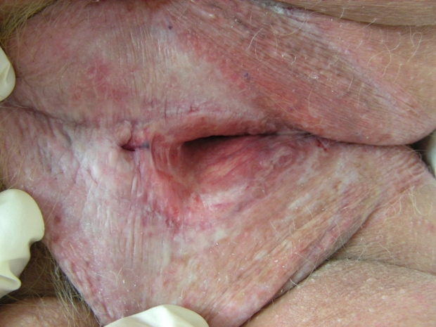
Lichen sclerosis
- Vaginal Discharge --> This case shows yeast vulvovaginitis – common cause of vaginal itching and/or discharge, common in hospitalized patients, especially if getting antibiotics.
- Also note, bacterial vaginosis is another common cause of discharge and often we need a microscope to differentiate between the two.
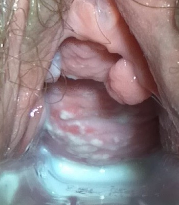
Yeast vulvovaginitis
- Trichomonas infection causing a “Strawberry cervix”
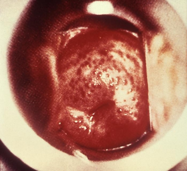
Trichomonas infection
Bimanual Exam
Pelvic Exam
Click this link to jump to the bimanual exam section of the video.
Insert index finger and possibly additional finger (with lubricant) in vaginal canal with non-dominant had free to palpate the abdomen.
o Look for:
- Cervical motion tenderness – could suggest cervicitis or pelvic inflammatory disease
- Position of uterus (image in folder) – helpful to know if you may not be able to palpate uterus if axial or retroflexed
- Adnexal size and tenderness – looking for any adnexal masses.
- Obesity may create a challenge to appreciating the adnexa.
- The post-menopause ovary can often be too small to palpate.
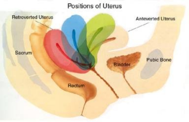
Position of uterus
Rectovaginal Exam
Pelvic Exam
Click here to jump to the rectovaginal exam section of the video.
The purpose of the exam is to:
- Appreciate a retroflexed uterus and adnexal masses
- Look for presence of hemorrhoids, polyps/growths and assess the tone of the rectal sphincter
Technique
- Place lubricant on index and middle finger of dominant hand then insert index finger into vaginal canal
- Insert middle finger into rectum, asking patient to bare down and insert fingers as far possible
- Appreciate
- Retroflexed uterus
- Nodularity along ureteral sacral ligament and cul-de-sac behind uterus – suggesting endometriosis
- To find ureteral sacral ligament, pull the cervix anterior with index finger to stretch and palpate for the ligament with finger inserted into rectum
- Adnexa – looking for masses and tenderness
- Rectal or anal masses/polyps
Special Considerations
Pelvic Exam
o Hospitalized patients – Given a proper exam table with leg rests is often not available, if possible delay this exam until patient can be seen in clinic. However if speculum exam is needed immediately, one method is to place an inverted bedpan under the patient’s buttocks to raise pelvis. If yeast vulvovaginitis is suspected, you or patient can obtain a swab and look under microscope or send to lab.
o Patient’s first exam or elderly patients – May require more discussion and education prior to the exam
o Patients with history of trauma – ensure patient knows they have control and can stop the exam at anytime
Key Learning Points
The key topics of the pelvic exam include:
- Understanding the types of speculums
- Knowling how to prepare your patient for the exam
- Palpation of the abdomen
- Vulvar and speculum examination
- The bimanual Exam
- The rectovaginal exam
- Performng the exam in the hosptialized patient (without an exam table)
Related to Pelvic Exam
- Hip Region Exam, Approach to
- Breast Exam
- Rectal Exam
The Stanford Medicine 25
- Aortic Regurgitation Exam
- Ankle Brachial Index
- Ankle and Foot Exam
- Ascites & Venous Patterns
- Bedside Ultrasound
- Breast Exam
- Cardiac Second Sounds
- Carpal Tunnel Exam
- Cerebellar Exam
- Deep Tendon Reflexes
- Dermatology Exam: Acne vs. Rosacea
- Dermatology Exam: Learning the Language
- Dermatology Exam: Nevi (Mole) Exam
- Fundoscopic Exam (Ophthalmoscopy)
- Gait Abnormalities
- Hand Exam
- Hip Region Exam
- Internal Capsule Stroke
- Involuntary Movements and Tremor Diagnosis: Types, Causes, and Examples
- Knee Exam
- Liver Exam
- Low Back Exam
- Lymph Node Exam
- Neck Vein Exam
- Pelvic Exam
- Precordial Movements in the Cardiac Exam
- Pulmonary Exam: Percussion & Inspection
- Pupillary Responses
- Pulsus Paradoxus and Blood Pressure Measurement Techniques
- Rectal Exam
- Spleen Exam
- Tarsal Tunnel Exam
- Thyroid Exam
- Tongue Exam
- Liver Disease, Head to Foot
- Visit the 25
- Shoulder Exam Tutorial
- Parkinson's Disease Exam
- Diastolic Murmurs Exam
- Dermatology Exam: Nevi (Mole) Exam