Bedside Ultrasound Examination
Bedside ultrasound has become a standard part of the exam for many clinicians. Here we review some basic aspects of the bedside exam and the ultrasound machines used.
Introduction to Bedside Ultrasound
The portability and availability of ultrasound is rapidly advancing bedside examination in places like the emergency room, where the FAST (Focused Assessment Sonography in Trauma) exam is becoming the standard of care. Bedside, or point of care, ultrasound has not yet become standardized for internists, but its utility cannot be ignored. We believe that teaching bedside ultrasound to the next generation of internists has the potential to standardize its use while bringing the internist back to the bedside.
Bedside ultrasound is not a replacement for sonographic studies performed by radiologists or cardiologists. Bedside ultrasound should be used to answer specific questions in real time. An example is the increasingly common use of bedside ultrasound by intensivists to estimate left ventricular function, an estimation not easily assessed by other physical exam maneuvers. The intensivist is able to answer a specific question in a timely fashion without ordering a complete echocardiogram. As bedside ultrasound is becoming more common it will be important that we educate patients to this difference, lest patients misunderstand the exams that are performed.
Currently there is no one clear model for teaching bedside ultrasound, with different institutions across the world creating their own approaches. We anticipate a steep learning curve especially when practicing on healthy patients. Sick patients, on the other hand, represent greater challenges and we feel that it will be important for residents to learn at both levels.
Ultrasound Machines
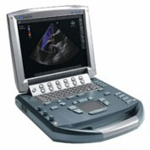
M-Turbo
These are easy to bring to the bedside and popular in the ER and ICU settings. They are durable and easy to use while retaining many powerful features.
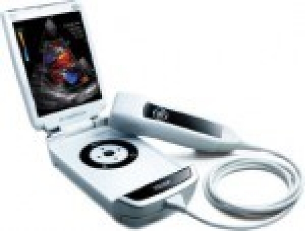
Hand Held Vscan
A truly pocket sized ultrasound that seems ready to bring ultrasound to the bedside of every patient by putting ultrasound in the pocket of physicians.
Probes
Ultrasound probes come in many different types and for our purposes we should be familiar with three different types.
Linear Probe
A linear probe uses high frequency ultrasound to create high resolution images of structures near the body surface. This makes the probe ideal for vascular imaging and certain procedures such as central line placement.
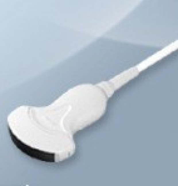
Curvilinear Probe
A curvilinear probe uses lower frequency ultrasound allowing a deep penetration and a wide depth of field, which is excellent for viewing intra-abdominal structures.
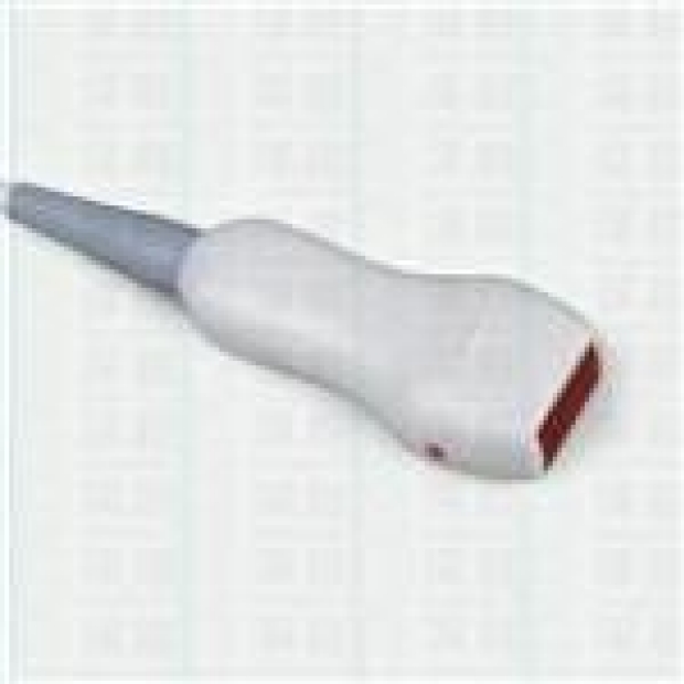
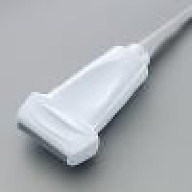
Phased Array Probe
Phased array probes give a large depth of field with a small footprint allowing the ultrasound to view deep structures though a small acoustic window. This makes it the ideal probe for viewing structure in the chest as the ultrasound waves are beamed between the ribs.
Key Learning Points
- Introduction to the ultrasound machines (used at Stanford Medical Center)
- Ultrasound Basics
Related to Examination of the Liver
The Stanford Medicine 25
- Aortic Regurgitation Exam
- Ankle Brachial Index
- Ankle and Foot Exam
- Ascites & Venous Patterns
- Bedside Ultrasound
- Breast Exam
- Cardiac Second Sounds
- Carpal Tunnel Exam
- Cerebellar Exam
- Deep Tendon Reflexes
- Dermatology Exam: Acne vs. Rosacea
- Dermatology Exam: Learning the Language
- Dermatology Exam: Nevi (Mole) Exam
- Fundoscopic Exam (Ophthalmoscopy)
- Gait Abnormalities
- Hand Exam
- Hip Region Exam
- Internal Capsule Stroke
- Involuntary Movements and Tremor Diagnosis: Types, Causes, and Examples
- Knee Exam
- Liver Exam
- Low Back Exam
- Lymph Node Exam
- Neck Vein Exam
- Pelvic Exam
- Precordial Movements in the Cardiac Exam
- Pulmonary Exam: Percussion & Inspection
- Pupillary Responses
- Pulsus Paradoxus and Blood Pressure Measurement Techniques
- Rectal Exam
- Spleen Exam
- Tarsal Tunnel Exam
- Thyroid Exam
- Tongue Exam
- Liver Disease, Head to Foot
- Visit the 25
- Shoulder Exam Tutorial
- Parkinson's Disease Exam
- Diastolic Murmurs Exam
- Dermatology Exam: Nevi (Mole) Exam